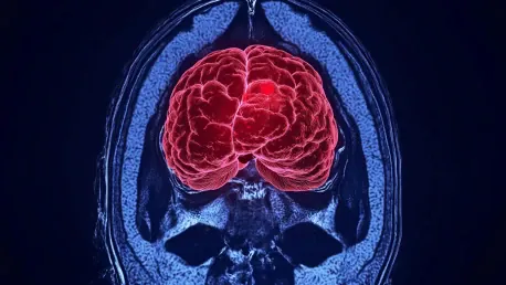
GE Healthcare has made a notable breakthrough in the field of medical imaging by developing an advanced Artificial Intelligence (AI) model that leverages Amazon Web Services (AWS) to enhance the interpretation of MRI images. This innovative AI model marks a substantial advancement in medical imaging, particularly for MRI scans, which have historically been complex and data-intensive. MRI images are inherently intricate and involve substantial data, making traditional analysis methods labor-intensive and limited in scope. Historically, developers working with large language models (LLMs) for MRI data analysis had to simplify these complex images by slicing them into 2D segments. This approach merely approximated the original 3D structure of the scans, thereby hindering the diagnostic capability of AI models, particularly in cases involving detailed anatomical structures or severe medical conditions like brain tumors, skeletal disorders, or cardiovascular diseases.
The Breakthrough: Full-Body 3D MRI Research Foundation Model
Overcoming Traditional Limitations
Recognizing these limitations, GE Healthcare has successfully introduced the industry’s first full-body 3D MRI research foundation model (FM). Presented at AWS re:Invent, this model allows for the utilization of comprehensive 3D images, providing an unprecedented level of detail and fidelity in medical imaging. The foundation model was meticulously developed using AWS’s robust infrastructure, handling over 173,000 images from more than 19,000 individual studies. Remarkably, the model required only a fifth of the computational power traditionally needed for similar tasks. This significant reduction in computational power means that the foundation model is more accessible and efficient, addressing one of the primary barriers in medical imaging advancements. By enabling the use of comprehensive 3D images, the model enhances the depth and precision of medical diagnostics, providing unprecedented insights into complex medical conditions. The transition from 2D to 3D imaging creates a paradigm shift, allowing for the visualization of anatomical structures in their entirety, which is particularly crucial for diagnosing intricate conditions such as brain tumors, vascular diseases, and musculoskeletal disorders.
Early Evaluation and Vision
Currently, the model remains in an evolutionary research phase and is not yet commercialized. Mass General Brigham is among the earliest evaluators set to begin experimenting with this pioneering technology soon. GE HealthCare’s chief AI officer, Parry Bhatia, articulated the vision behind this development, emphasizing the aim to arm healthcare technical teams with powerful tools to expedite research and clinical applications more efficiently and cost-effectively. The vision articulated by Bhatia underscores the potential of this AI model to revolutionize the field of MRI imaging by making advanced diagnostic tools more accessible to healthcare providers worldwide. By reducing the resource requirements for 3D MRI analysis, this model paves the way for broader adoption and integration of advanced imaging techniques in clinical settings. This can lead to faster diagnosis and treatment planning, significantly improving patient outcomes across various medical disciplines. Moreover, the partnership with AWS ensures that the infrastructure supporting this model remains scalable, secure, and compliant with regulatory standards, fostering innovation in medical technology.
Real-Time Analysis and Medical Procedures
Enhancing Medical Procedures
A notable capability of the new model lies in its ability to enable real-time analysis of intricate 3D MRI data. This advancement holds considerable potential to enhance medical procedures, such as biopsies, radiation therapy, and robotic surgery, by offering more precise and immediate data interpretation. The real-time capability means that clinicians can make informed decisions swiftly, which is critical in time-sensitive medical situations. The ability to perform real-time analysis significantly enhances the precision and efficiency of interventional procedures. For instance, during biopsies, accurate and immediate interpretation of MRI data can guide the needle to the target area with greater accuracy, reducing the risk of complications and improving diagnostic yield. In radiation therapy, real-time imaging allows for more precise targeting of tumors, sparing healthy tissue and enhancing treatment efficacy. Robotic surgery stands to benefit immensely, as the model’s real-time data interpretation capabilities can support complex surgical maneuvers, improve surgical precision, and reduce operative times, ultimately contributing to better patient outcomes and faster recovery.
Leveraging Generative AI and LLMs
Generative AI and LLMs are not novel territories for GE Healthcare. The team has been leveraging advanced technologies for over a decade. One of GE Healthcare’s flagship products is AIR Recon DL, a deep learning-based reconstruction algorithm designed to accelerate the acquisition of crisp, clear images by radiologists. This algorithm strategically removes noise from raw images, improves the signal-to-noise ratio, and reduces scan times by up to 50%. Since its launch in 2020, AIR Recon DL has effectively scanned 34 million patients. By leveraging generative AI, GE Healthcare continuously innovates to enhance the capabilities of its imaging solutions. The successful integration of AIR Recon DL into clinical practice is a testament to the company’s expertise in deploying AI-driven technologies that improve the accuracy and efficiency of medical imaging. With the development of the full-body 3D MRI research foundation model, GE Healthcare builds on this legacy, pushing the boundaries of what is possible in diagnostic imaging. The company’s decade-long experience with advanced AI technologies positions it at the forefront of medical innovation, capable of delivering solutions that significantly enhance patient care and clinical outcomes.
Development Journey and Model Capabilities
Multimodal Model Design
The development journey for GE Healthcare’s MRI foundation model began in early 2024. Designed as a multimodal model, it supports various functions such as image-to-text searching, linking images and words, and disease segmentation and classification. This enables healthcare professionals to derive more comprehensive details from a single scan, thereby facilitating faster and more accurate diagnosis and treatment. The multimodal nature of the model represents a significant leap forward in medical imaging technology. By integrating various data types—such as images and textual descriptions—into a unified framework, the model enhances the diagnostic process. Image-to-text searching allows clinicians to find relevant case studies and medical literature linked directly to MRI images, providing valuable context and aiding in the interpretation of complex cases. Disease segmentation and classification capabilities streamline the diagnostic workflow, enabling faster identification of pathologies and more targeted treatment planning. This holistic approach to medical imaging ensures that clinicians have access to all pertinent information, leading to more informed decision-making and improved patient outcomes.
Promising Early Results
The early results have been promising, with the model outperforming other publicly available research models in tasks like the classification of prostate cancer and Alzheimer’s disease. It has also shown significant improvement in matching MRI scans with text descriptions in image retrieval, with accuracy rates jumping to 30% compared to a mere 3% by similar models. These impressive early results indicate the model’s potential to set new standards in medical imaging. In the classification of conditions such as prostate cancer and Alzheimer’s disease, the model’s enhanced accuracy aids in early detection and intervention, which are critical for successful treatment outcomes. The significant improvement in image retrieval demonstrates the model’s ability to provide clinicians with relevant information effectively, further supporting diagnosis and treatment planning. As the model continues to evolve, these capabilities are expected to expand, offering even greater precision and utility across a broader range of medical applications. The high accuracy rates achieved by the model underscore its potential to revolutionize diagnostic imaging, paving the way for more personalized and effective healthcare solutions.
Training and Data Handling Techniques
Specialized Imaging Techniques
Training the model for MRI data analysis requires particular types of datasets to support different imaging techniques. For instance, the T1-weighted imaging technique focuses on highlighting fatty tissue while decreasing water signal, and the T2-weighted imaging technique enhances water signal. Together, these methods provide a comprehensive image that aids clinicians in detecting abnormalities such as tumors, trauma, or cancer. These specialized imaging techniques are crucial for providing a detailed and accurate visualization of various tissue types and anatomical structures. T1-weighted images are particularly effective for identifying structures rich in fat, such as bone marrow and certain brain regions, making them invaluable in diagnosing conditions like bone fractures and tumors. On the other hand, T2-weighted images excel in highlighting areas with high water content, such as edema or inflammation, which are often indicative of trauma or infection. By leveraging both techniques, the model can deliver a more comprehensive and nuanced picture of the patient’s condition, enhancing the accuracy of diagnoses and guiding more targeted treatment strategies.
Handling Diverse and Incomplete Datasets
To navigate the challenges of diverse and sometimes incomplete datasets, GE Healthcare developers employed a “resize and adapt” strategy, allowing the model to process and respond to variations in the data. They further trained the model to ignore missing data and focus on the available information effectively. These methods were akin to solving a complex puzzle with some missing pieces, ensuring that the model could deliver accurate results even under imperfect conditions. The “resize and adapt” strategy is crucial for accommodating the variability inherent in medical imaging datasets. Medical images can vary significantly in quality, resolution, and completeness due to differences in scanning equipment, patient positioning, and other factors. By training the model to adapt to these variations, GE Healthcare ensures that it remains robust and reliable across a wide range of scenarios. The ability to ignore missing data and focus on available information enhances the model’s resilience, ensuring that it can provide accurate and clinically relevant insights even when faced with less-than-ideal input. This adaptability is essential for delivering consistent performance in real-world clinical settings, where variability is a constant challenge.
Advanced Learning Techniques and Multimodality
Semi-Supervised Learning
Additionally, semi-supervised student-teacher learning techniques were employed, particularly useful in scenarios with limited data. This method involves two neural networks: one (the teacher) creates labels that assist the other (the student) in learning and predicting future labels. These self-supervised technologies reduce the dependency on large labeled datasets, enabling the model to learn more efficiently from raw images. Semi-supervised learning techniques are particularly valuable in medical imaging, where labeled datasets can be scarce and expensive to obtain. By leveraging self-supervised technologies, the model can effectively utilize available data, enhancing its learning capabilities and reducing the need for extensive manual annotation. The student-teacher framework facilitates the transfer of knowledge within the model, enabling it to make more accurate predictions and interpretations even with limited labeled data. This approach not only accelerates the development process but also enhances the model’s ability to adapt to new and evolving medical imaging techniques, ensuring its long-term relevance and efficacy in clinical practice.
Multimodal Integration
The model’s multimodality stands out as a significant advancement. Traditional models were often unimodal, focusing only on images or text separately. The new model can transition seamlessly from image to text and vice versa, integrating various data types into a unified workflow. GE Healthcare emphasizes adherence to compliance standards and careful use of de-identified datasets to navigate privacy concerns effectively. By integrating multiple data types, the model provides a more holistic view of the patient’s condition, facilitating comprehensive and accurate diagnoses. The seamless transition between image and text data enhances the clinical workflow, allowing healthcare providers to access all relevant information within a single platform. This integration reduces the need for cross-referencing disparate sources, saving time and reducing the risk of errors. GE Healthcare’s commitment to compliance and privacy ensures that the model’s adoption aligns with regulatory standards, safeguarding patient data and maintaining trust. This comprehensive approach to multimodal integration represents a significant step forward in the application of AI in medical imaging, promising to enhance diagnostic accuracy and improve patient outcomes across various medical fields.


